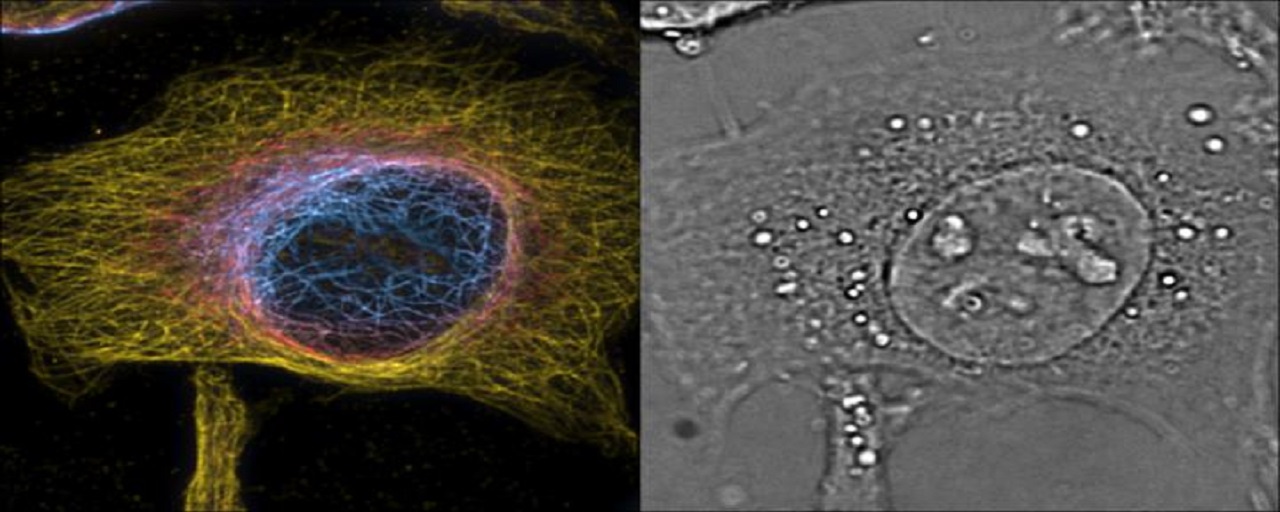
Super-resolution microscopy has been known to have the ability to capture a high-quality view of interior structures and organelles in cells. However, it is severely restricted, because this only works if examining something that remains static. Living cells would require a much more complex way to be able to study them as thoroughly. Luckily, a team led by Professor Theo Lasser, the head of the Laboratory of Biomedical Optics (LOB) have developed a way to do this.
Super-resolution microscopy has been known to have the ability to capture a high-quality view of interior structures and organelles in cells. However, it is severely restricted, because this only works if examining something that remains static. Living cells would require a much more complex way to be able to study them as thoroughly. Luckily, a team led by Professor Theo Lasser, the head of the Laboratory of Biomedical Optics (LOB) have developed a way to do this. They have combined the sensitivity and high time-resolution of phase imaging with the specificity and high spatial resolution of fluorescence microscopy in order to visualize cells in 3D. To read more, check out the full article.
Filed Under: Rapid prototyping


