Electromagnetic radiation with a frequency between 3 × 10¹⁶ Hz (30 petahertz) and 3 × 10¹⁹ Hz (30 exahertz) falls into the X-ray category. The wavelengths are shorter than ultraviolet and longer than gamma radiation.
Wilhelm Röntgen was the first to describe in detail this radiation, which he termed X-rays because they had been previously unknown. In recognition, he was awarded the first Nobel Prize and also an honorary medical degree.
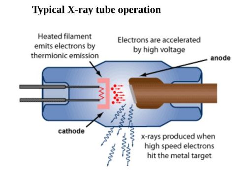 X-rays are produced when high-energy charged particles, electrons or ions, strike a material. A common method for generating X-rays makes use of a vacuum tube. A high voltage accelerates electrons emitted from a hot cathode to a high velocity. Focused into a narrow beam, the electrons strike an anode, which emits the X-rays. The target for medical X-rays is tungsten or a rhenium alloy thereof, or molybdenum. A copper anode is used in crystallography applications, and the anode is cobalt when the target contains iron, which otherwise would fluoresce. Another situation in which X-rays are produced is when electrons make transitions between lower energy levels in atoms of heavy elements.
 It takes a high voltage to produce X-radiation of sufficient energy for practical applications, typically 80 kV. X-rays generated as described are confined to specific, discrete frequencies which depend upon the anode material.
Another type of emission is known as Bremsstrahlung. This radiation, produced from electron scattering by a strong electric field, manifests as continuous X-ray emission.
Besides medical and dental applications, X-rays have numerous uses, ranging from X-ray crystallography to airport security.
High-energy astronomical objects have been found to be powerful X-ray sources. X-rays emitted by astronomical objects cannot be received on the earth’s surface because the atmosphere is opaque to these frequencies (fortunately for life on earth). Balloons and rockets have been used, but orbiting satellites have made possible a new epoch in X-ray astronomy.
X-ray emission from the sun has been noted for over 50 years, but currently, levels hundreds of thousands of times greater than the sun’s total emission are measured from numerous deep-sky objects. What creates such massive emission is high energy, particularly involving neutron stars including pulsars, and black holes. Of course, black holes do not emit X-rays except as a tiny amount of Hawking radiation, but matter just outside the event horizon, subjected to incredible amounts of energy, emits huge amounts of X-radiation before being silenced forever.
The big player in X-ray astronomy is the Chandra X-Ray Observatory (CXO), launched by NASA in 1999. Its planned lifespan was five years, but remarkably it is going strong after 16 years.
X-rays are a kind of ionizing radiation. As a quick review of the concept, when the number of electrons orbiting an atom is equal to the number of protons, the atom has neither a positive nor a negative charge. It is electrically neutral. Under certain circumstances, an atom will lose an electron from its outer shell. Then, the atom is no longer neutral, but instead it has a positive charge. That is, it has become a positive ion. There are several consequences, some good and some not so good. For living beings, the biological material is altered, the end point being that it cannot sustain life.
One way atoms lose electrons is from exposure to ionizing radiation, as in atom bomb. Exposure can cause various degrees of radiation sickness and genetic mutations that years later may manifest as malignant tumors. When a sufficiently intense beam of X-rays strikes matter, electrons are blasted out of the outer valence shell so neutral atoms become charged ions. In the process, molecular bonds are disrupted and the matter is abruptly transformed. Needless to say, this has profound biological implications. Living tissue is altered in a variety of ways most of which preclude continued life. Cells can be outright destroyed or genetically modified so that they become cancerous.
Non-ionizing radiation does not affect biological material in this way. But it can still be damaging and even lethal if it is intense enough, only because it makes for temperature rise.
Some materials can be ionized by being exposed to voltage. This is a function of the level of voltage and the duration. When a material is ionized, it becomes more conductive.
Lightning, with characteristic zigzag paths, is caused by ionization of the air, which then becomes highly conductive. A continuous path is created and there is sudden current flow. Ionization also happens in fluorescent lamps and in the resonant cavities of lasers. Other examples are when insulation associated with an electrical conductor breaks down from voltage fatigue, or when an automotive spark plug fires.
Soon after the first X-ray tube was built, applications were found in the field of medicine. X-rays pass through the human body more readily through soft tissue than bones, which are denser. Because X-rays affect photographic film in the same way as visible light, it is possible to create images of bone fractures and breaks. Similarly, cancerous masses are denser than healthy tissue and can be imaged for diagnostic purposes. The intensity and duration of the X-ray exposure must be carefully calibrated so the procedure does not do more harm than good.
In addition to its use as a diagnostic procedure, X-rays are successful in a certain proportion of cases as anti-cancer therapy. Here, a much higher dose of X-radiation is involved compared to that in medical imaging procedures. Some patients are sickened and even killed by radiation therapy though a large number of individuals are saved. Before treatment, there is a careful risk assessment and dosage calibration that involves adjusting radiation intensity, duration, and number and spacing of exposures.
Multiple shaped radiation beams are aimed at different angles so as to intersect at the tumor. The object is to hit the malignant site with a larger dose compared to the surrounding healthy tissue. Radiation therapy is highly successful in destroying residual cancerous cells that may remain after surgery. The procedure is commonly used in conjunction with surgery, chemotherapy, hormone therapy and immunotherapy.
The post X-rays and ionizing radiation appeared first on Test & Measurement Tips.
![]()
Filed Under: Test & Measurement Tips